第3期 | Countstar与科研之间的故事
发布日期:2021-06-14 09:12:00浏览次数:2567
本周CountStar在最新的应用发表文献中选取了6篇分享给大家。第1篇是关于COVID-19单细胞测序相关的研究,文章通过对感染者和健康者的免疫细胞的转录谱测定,揭示了发病是机体免疫反应的动态性质。第3篇文章介绍了LncRNA-DILA1对于乳腺癌的调控机理。第4篇文章在免疫治疗领域新型化学材料的应用前景探究。
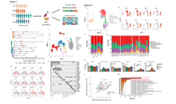
Abstract :In COVID-19 caused bySARS-CoV-2 infection, the relationship between disease severity and the hostimmune response is not fully understood. Here we performed single-cell RNAsequencing in peripheral blood samples of five healthy donors and 13 COVID-19patients including moderate, severe and convalescent cases. Through determiningthe transcriptional profiles of immune cells, coupled with assembled T cellreceptor and B cell receptor sequences, we analyzed the functional propertiesof immune cells. Most cell types in COVID-19 patients showed a stronginterferon-alpha response, and an overall acute inflammatory response.Moreover, intensive expansion of highly cytotoxic effector T cell subsets, suchas CD4+ Effector-GNLY (Granulysin), CD8+ Effector-GNLY and NKT CD160, wasassociated with convalescence in moderate patients. In severe patients, theimmune landscape featured a deranged interferon response, profound immuneexhaustion with skewed T cell receptor repertoire and broad T cell expansion.These findings illustrate the dynamic nature of immune responses during thedisease progression.
Abstract:Heat stress induced by continuous high ambient temperaturesor strenuous exercise in humans and animals leads to intestinal epithelialdamage through the induction of intracellular stress response. However, theprecise mechanisms involved in the regulation of intestinal epithelial cellinjury, especially intestinal stem cells (ISCs), remain unclear. Thereby, invitro a confluent monolayer of IPEC‐J2 cells was exposed to the high temperatures (39, 40,and 41°C), the IPEC‐J2 cell proliferation, apoptosis, differentiation, andbarrier were determined, as well as the expression of GRP78, which is a markerprotein of endoplasmic reticulum stress (ERS). The Wnt/β‐catenin pathway‐ mediated regenerative response was validated using R‐spondin 1(Rspo1). And ex‐vivo, three‐dimensional cultured enteroids were developed from pigletjejunal crypt and employed to assess the ISC activity under heat exposure. Theresults showed that exposure to 41°C for 72 hr, rather than 39°C and 40°C,decreased IPEC‐J2 cell viability, inhibited cell proliferation anddifferentiation, induced ERS and cell apoptosis, damaged barrier function andrestricted the Wnt/β‐cateninpathway. Nevertheless, Wnt/β‐cateninreactivation via Rspo1 protects the intestinal epithelium from heat exposure‐inducedinjury. Furthermore, exposure to 41°C for 24 hr reduced ISC activity,stimulated crypt‐cell apoptosis, upregulated the expression of GRP78 andcaspase‐3, and downregulated the expression of β‐catenin, Lgr5, Bmi1, Ki67, KRT20, ZO‐1,occludin, and claudin‐1. Taken together, we conclude that heat exposure inducesERS and downregulates the Wnt/β‐cateninsignaling pathway to disrupt epithelial integrity by inhibiting the intestinalepithelial cell proliferation and stem cell expansion.
[Nat Commun] IF=12.121 PMID: 33139730
Abstract:Cyclin D1 is one of the most important oncoproteins thatdrives cancer cell proliferation and associates with tamoxifen resistance inbreast cancer. Here, we identify a lncRNA, DILA1, which interacts with CyclinD1 and is overexpressed in tamoxifen-resistant breast cancer cells.Mechanistically, DILA1 inhibits the phosphorylation of Cyclin D1 at Thr286 bydirectly interacting with Thr286 and blocking its subsequent degradation,leading to overexpressed Cyclin D1 protein in breast cancer. Knocking downDILA1 decreases Cyclin D1 protein expression, inhibits cancer cell growth andrestores tamoxifen sensitivity both in vitro and in vivo. High expression ofDILA1 is associated with overexpressed Cyclin D1 protein and poor prognosis inbreast cancer patients who received tamoxifen treatment. This study shows the previouslyunappreciated importance of post-translational dysregulation of Cyclin D1 con tributingto tamoxifen resistance in breast cancer. Moreover, it reveals the novelmechanism of DILA1 in regulating Cyclin D1 protein stability and suggests DILA1is a specifific therapeutic target to downregulate Cyclin D1 protein andreverse tamoxifen resistance in treating breast cancer.
Abstract:Mounting evidence suggests that immunotherapies are apromising new class of anticancer therapies. However, the immunosuppressivetumor microenvironment (TME), poor immunogenicity, and off-target toxicityhinder the broader implementation of immunotherapies. Here, we describe a novelstrategy combining chemotherapy and immunotherapy to modulate the TME bysystemically and concurrently delivering the chemotherapeutic agent SN38(7-ethyl-10-hydroxycamptothecin) and the STING agonist DMXAA(5,6-dimethylxanthenone-4-acetic acid) into tumors using triblock copolymernanoparticles, named PS3D1@DMXAA, which enhances antigen cross-presentation andinduces the conversion of the immunosuppressive TME to immunogenic TME throughthe newly found synergistic function between SN38 and STING activation.PS3D1@DMXAA thus shows potent therapeutic efficacy in three mice tumor modelsand elicits remarkable therapeutic benefit when combined with anti–PD-1therapy. Our engineered nanosystem offers a rational design of an effectiveimmunotherapy combination regimen to convert uninflamed “cold” tumors into“hot” tumors, addressing the major challenges immunotherapies faced.
5.Intrinsic ColorSensing System Allows for Real-Time Observable Functional Changes on Human InducedPluripotent Stem Cell-Derived Cardiomyocytes
固有颜色传感系统允许实时观察到人类诱导的多能干细胞衍生心肌细胞的功能变化
[ACS Nano] IF=14.588 PMID: 32609489
Abstract:Stem-cell based in vitro difffferentiation for diseasemodeling offffers great value to explore the molecular and functionalunderpinnings driving many types of cardiomyopathy and congenital heartdiseases. Nevertheless, one major caveat in the application of in vitrodifffferentiation of human induced pluripotent stem cell (hiPSC)-derivedcardiomyocytes (hiPSC-CMs) involves the immature phenotype of the CMs. Most ofthe existing methods need complex apparatus and require laborious procedures inorder to monitor the cardiac difffferentiation/maturation process and oftenresult in cell death. Here we developed an intrinsic color sensing systemutilizing a microgroove structural color methacrylated gelatin fifilm, whichallows us to monitor the cardiac difffferentiation process of hiPSC-derived cardiac progenitorcells in real time. Subsequently this system can be employed as an assay systemto live monitor induced functional changes on hiPSC-CMs stemming from drugtreatment, the effffects of which are simply revealed through color diversity.Our research shows that early intervention of cardiac difffferentiation throughsimple physical cues can enhance cardiac difffferentiation and maturation tosome extent. Our system also simplififies the previous complex experimentalprocesses for evaluating the physiological effffects of successfuldifffferentiation and drug treatment and lays a solid foundation for futuretransformational applications.
Countstar Fluorescence Cell Analyzer
- 上一篇:合规?还是不合规?得看这几点!
- 下一篇:微载体培养的细胞计数 第二期